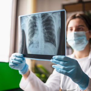
Cardiovascular Imaging
Cardiovascular imaging services play a crucial role in diagnosing and treating various medical conditions related to your heart. We provide several imaging techniques to deliver the best diagnostic information to our patients, such as:
- Cardiac CT
- Cardiac MRI
- Echocardiography
- Nuclear imaging
Cardiac CT
Our modern CT scanners provide detailed information about your heart's structure and function by taking high-resolution 3D images. This is a non-invasive method of taking multiple X-rays of your heart and vascular system. It can also provide data about the anatomy of the coronary arteries and great vessels, indicating blockages or narrowing.
The speed of the scanner allows imaging within a few seconds. The study requires special computer processing and analysis. We interpret all cardiac CT studies performed at Bellin Hospital. All X-ray studies expose patients to some radiation. As scanners have improved, the dose to patients has reduced. However, we consider radiation exposure as part of the minimal risk of the study.
Cardiac MRI
Cardiac MRI imaging uses a powerful magnetic field, radio frequency pulses and a computer to produce detailed pictures of the heart. Magnetic resonance imaging is a non-invasive test that helps providers diagnose and treat medical conditions. It does this without using any radiation.
Performing cardiac MRI helps:
- Evaluate the anatomy and function of the heart, valves, major vessels and surrounding structures.
- Diagnose and manage a variety of cardiovascular problems.
- Detect and evaluate the effects of coronary artery disease.
- Plan a patient's treatment for cardiovascular problems and monitor a patient's progress.
Echocardiography
Echocardiography is a non-invasive imaging test of the heart that uses ultrasound waves to acquire pictures. There is no radiation risk for this kind of test. This information gives a detailed analysis of the structure and function of the heart.
There are two types of echocardiograms:
- Transthoracic echocardiogram (TTE)
- Transesophageal echocardiography (TEE)
A transthoracic echocardiogram, or TTE, is a standard heart ultrasound test done in the office. The imaging process captures the heart from various positions. This test takes approximately 60 minutes to complete. There is no preparation for it.
Transesophageal echocardiography, or TEE, uses ultrasound technology to provide imaging of the heart structures that surface echocardiography doesn't typically show. This is because the sound waves travel a shorter distance. They don't need to pass through the skin, chest, muscle or bone. TEE uses a probe in the esophagus to get a clear image of the heart. Patients that need more detailed information and imaging should follow this recommendation.
What to Expect for Your TEE
Do not eat or drink anything except small sips of water for at least eight hours before the test. Tell your provider if you have any allergies or sensitivities to medications.
- First, we will place electrodes on your chest and administer sedation to relax you.
- Once you relax, we will pass the probe and proceed with cardiac imaging.
- The entire test takes approximately 60 minutes, with an additional hour to recover.
- Shortly after your recovery time is over, you may go home.
- Due to sedation, please arrange for someone to drive you home.
Nuclear Imaging
Stress testing checks blood flow to the heart to find blockages in the arteries that supply the heart. Our providers do this to evaluate symptoms or to work up certain heart conditions. When patients cannot walk on a treadmill, we administer medication through an IV to stress the heart.
We use a nuclear stress test to determine how well your heart works during physical activity and at rest. This testing involves a particular substance given through an IV that becomes visible on images taken by a special camera. It examines blood flow to the heart muscle and identifies any areas of damage.
